Unveiling Neck Anatomy: Structure, Muscles, & Essential Limits
The human neck, a marvel of biological engineering, serves as the critical bridge connecting the head to the torso and upper limbs. Far more than just a support column, it is a complex conduit for vital structures, including the central nervous system, major blood vessels, and the pathways for breathing and digestion. Understanding the intricate Anatomia Del Cuello – or the anatomy of the neck – is fundamental, whether for medical professionals, fitness enthusiasts, or anyone curious about the human body. Its unique design allows for an incredible range of motion while simultaneously protecting delicate internal components, making it a focal point of both strength and vulnerability.
Typically cylindroid in shape, the neck's circumference broadens at its base, where it merges with the shoulders and chest. Its length is primarily defined by the stacking of the seven cervical vertebrae (C1-C7), each playing a unique role, from the atlas (C1) supporting the skull to the axis (C2) facilitating head rotation. The thickness or width of the neck, however, is a more variable characteristic, influenced significantly by the development of its numerous muscle masses and the distribution of adipose tissue. Factors such as age, sex, body mass index, and even individual genetics contribute to the variations in neck morphology, highlighting its dynamic nature.
Defining the Borders: A Comprehensive Guide to Neck Limits
Pinpointing the exact boundaries of the neck is crucial for anatomical studies, surgical procedures, and diagnostic imaging. These imaginary lines define where the head ends and the neck begins, and where the neck transitions into the chest and upper extremities.
Superior Limits of the Neck
The superior boundary of the neck is marked by a comprehensive line that encircles the head. Anteriorly, it follows the inferior border of the mandible, an area rich in structures such as the submaxillary gland, submental and submandibular lymph nodes, the facial artery and vein, and parts of the intermaxilloparotid fascia. Posteriorly, this limit sweeps from the chin to the external occipital protuberance at the back of the skull. Along its lateral path, it encompasses the angle of the jaw and extends to the mastoid process of the temporal bone, located just behind the ear. This line also conceptually includes the superior nuchal line and a horizontal projection from the temporomaxillary joint to the external occipital protuberance, providing a robust definition for this crucial interface.
Inferior Limits of the Neck
The inferior boundary represents the transition from the neck to the thorax and upper limbs. It is defined by the superior surface of the clavicles, the collarbones, and continues medially along the superior border of the manubrium of the sternum. Posteriorly, this line extends between the acromioclavicular joints on either side, connecting them to the spinous process of the seventh cervical vertebra (C7). This comprehensive delineation ensures that the structures within this transitional zone are correctly identified as belonging to the neck.
Anterior and Posterior Limits
The anterior limit of the neck is less about a single bone and more about fascial and muscular confluence. It's essentially formed by the convergence of the common fascia, often referred to as the infrahyoid white line, and the fascias of the pre-laryngeal muscles. This area is central to the throat and houses several vital structures related to speech, swallowing, and breathing. The posterior limit, conversely, is marked by the convergence of prominent muscles: the sternocleidomastoid, the splenius capitis, and the levator scapulae. An important landmark in this region is Erb's point, where the greater auricular nerve emerges, making it a significant clinical reference point for nerve blocks and surgical considerations.
Just inferior to Erb's point lies the supraclavicular triangle, a critical anatomical space. Its boundaries are defined anteriorly by the sternocleidomastoid muscle, posteriorly by the trapezius muscle, and inferiorly by the omohyoid muscle and the clavicle. Deep within this triangle lie the scalene muscles and the neurovascular sheath, highlighting its importance as a passageway for structures supplying the upper limb and neck.
The Muscular Tapestry: Regions and Key Muscles of the Neck
The muscles of the neck are responsible for its incredible mobility, posture, and protective functions. They can be broadly categorized into three main regions, each with specialized roles. For a deeper dive into the specific roles and intricate interplay of these muscle groups, explore our dedicated article: The Neck's Complex Musculature: From Suprahyoids to Scalenes.
- Lateral Region: This area includes muscles like the platysma, a superficial muscle responsible for expressions and tensing the skin of the neck; the powerful sternocleidomastoid, which allows for head rotation and flexion; and the scalene muscles (anterior, middle, and posterior), crucial for neck flexion and elevating the first two ribs during forced inspiration. The rectus capitis lateralis also resides here, aiding in lateral flexion of the head.
- Anterior or Hyoid Region: These muscles are grouped by their relationship to the hyoid bone, a U-shaped bone that acts as an anchor for the tongue and larynx.
- Suprahyoid Muscles: Located above the hyoid, these include the digastric, stylohyoid, mylohyoid, and geniohyoid muscles. They primarily function in elevating the hyoid bone and the floor of the mouth, playing vital roles in swallowing and speech.
- Infrahyoid Muscles: Situated below the hyoid bone, this group comprises the sternohyoid, omohyoid, sternothyroid, and thyrohyoid muscles. Their main action is to depress the hyoid bone and larynx, assisting in vocalization and the latter stages of swallowing.
- Prevertebral Region: Deepest of all, these muscles lie directly in front of the cervical vertebrae. They include the rectus capitis anterior major and minor, and the longus colli. These muscles are key stabilizers of the head and cervical spine, facilitating subtle movements of the head and neck flexion.
The robust development of these muscle groups directly influences the structural integrity and appearance of the neck, offering both strength and flexibility to this critical region.
Beyond Muscles: Vital Structures and Protective Fascias
The neck is truly a crossroads, a vital passageway for an array of non-muscular structures essential for life. In an axial (horizontal) cross-section, one can appreciate the close proximity of the spinal cord and cervical vertebral column, illustrating the neck's role in protecting the central nervous system. This delicate balance of protection and passage highlights the neck's profound importance. For an in-depth exploration of how these intricate systems navigate this vital passage, refer to our article: Neck as a Crossroads: Vessels, Nerves, & Visceral Pathways.
Key Non-Muscular Structures:
- Arteries of the Neck: Major arteries like the carotid and vertebral arteries ascend through the neck, supplying blood to the brain and head.
- Veins of the Neck: The jugular veins, both internal and external, are prominent examples, responsible for draining deoxygenated blood from the head and neck back to the heart.
- Lymph Nodes of the Neck: Numerous lymph nodes are strategically scattered throughout the neck, forming a crucial part of the immune system, filtering lymph and defending against infection.
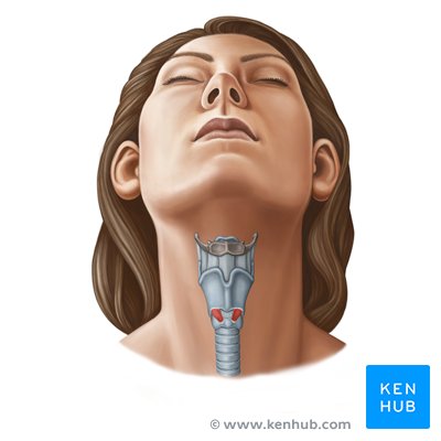
- Pharynx, Larynx, and Trachea: These visceral structures form the pathway for air (trachea, larynx) and food (pharynx, esophagus - which is just posterior to the trachea), enabling breathing, swallowing, and speech. The larynx, or voice box, is particularly complex, housing the vocal cords.
- Thyroid Gland: This endocrine gland, situated at the front of the neck, produces hormones critical for metabolism, growth, and development.
The Fascial System: Compartmentalization and Protection
The neck's various structures are not simply free-floating; they are meticulously organized and protected by a series of connective tissue layers known as fascias. These fascias serve multiple functions, including compartmentalizing structures, preventing the spread of infection, and providing planes for surgical dissection.
- Superficial Cervical Fascia: This outermost layer lies directly beneath the skin, enveloping the entire neck. It encases superficial muscles like the platysma, the sternocleidomastoid, the trapezius, and protects the superficial jugular vein.
- Middle Cervical Fascia (Pretracheal Fascia): This deeper layer surrounds the infrahyoid muscles and encloses the thyroid gland, trachea, and esophagus. It helps to delineate the central visceral compartment of the neck.
- Deep Cervical Fascia: This is the most complex of the fascial layers, with multiple divisions (investing layer, prevertebral layer, carotid sheath). It surrounds the deep muscles of the neck, the cervical vertebrae, and importantly, forms the carotid sheath which encases the common carotid artery, internal jugular vein, and vagus nerve. The prevertebral layer surrounds the vertebral column and its associated deep muscles, forming a strong protective barrier.
These fascial layers are vital for maintaining the structural integrity of the neck, providing both support and a degree of separation that is essential for both normal physiological function and surgical interventions.
Conclusion: The Neck – A Bridge of Life
The comprehensive understanding of the Anatomia Del Cuello reveals a region of unparalleled importance and complexity. From its bony framework of cervical vertebrae to its extensive muscular network, vital neurovascular bundles, and crucial visceral pathways, the neck performs an extraordinary array of functions. It supports the head, facilitates sensory input, enables communication, and serves as a literal bridge for all information and sustenance traveling between the brain and the body. Its intricate fascial system provides structural integrity and protection, yet also defines pathways for potential disease spread. Appreciating this anatomical masterpiece is key to grasping the full scope of human health, movement, and vulnerability. Maintaining neck health through good posture, ergonomic practices, and understanding its limits is paramount for overall well-being.