The neck, a relatively small but extraordinarily complex region, serves as the vital anatomical bridge connecting the head to the torso and upper limbs. Far from being a simple connector, it is a sophisticated crossroads, a bustling thoroughfare for a multitude of essential structures. Understanding the intricate Anatomia Del Cuello (anatomy of the neck) reveals a fascinating interplay of skeletal support, muscular action, and critical pathways for vessels, nerves, and visceral systems that are fundamental to human survival and function.
This dynamic region facilitates crucial movements of the head, provides protection for the spinal cord, and acts as a conduit for everything from the air we breathe to the signals that govern our entire body. Its cylindrical yet varied form adapts to age, sex, and individual body composition, typically spanning around 7-8 cm in length. What truly defines the neck's incredible importance, however, is the sheer concentration of life-sustaining elements packed within its confines.
The Neck: A Vital Anatomical Junction
At its core, the neck’s structure is anchored by the seven cervical vertebrae (C1-C7). These include the atypical C1 (atlas) and C2 (axis) that allow for extensive head movement, along with typical vertebrae C3-C6 and the transitional C7. The circumference of the neck is broader at its base, where it meets the shoulders and thorax, and its overall thickness is significantly influenced by the development of its numerous muscle masses and the distribution of adipose tissue.
Defining the Neck's Borders: A Topographical Guide
Pinpointing the exact limits of the neck is crucial for both anatomical study and clinical practice. These boundaries delineate the region and highlight its interconnectedness with adjacent body parts:
- Superior Limit: An imaginary line extends from the mentum (chin) anteriorly, along the inferior border of the mandible (where structures like the submandibular gland, submental and submandibular lymph nodes, and facial artery/vein reside), past the angle of the mandible, then posteriorly to the mastoid process of the temporal bone (behind the ear), and finally to the external occipital protuberance at the back of the skull. This superior boundary is also characterized by muscles like the stylohyoid, digastric, and the upper part of the sternocleidomastoid.
- Inferior Limit: This boundary is formed by the superior surface of the clavicles, extending laterally to the acromioclavicular joints. Anteriorly, it includes the superior border of the sternal manubrium, and posteriorly, it connects the acromial processes with the spinous process of the seventh cervical vertebra (C7).
- Anterior Limit: Defined by the convergence of the common fascia or the infrahyoid "white line" and the pre-laryngeal muscles.
- Posterior Limit: This region marks the confluence of significant muscles, including the sternocleidomastoid, splenius capitis, and levator scapulae. Clinically, the "Point of Erb" is a notable landmark here, where the greater auricular nerve emerges, making it relevant for nerve blocks and understanding sensory innervation. From Erb's point, extending downwards, lies the supraclavicular triangle, bounded anteriorly by the sternocleidomastoid, posteriorly by the trapezius, and inferiorly by the omohyoid muscle and clavicle. Deep within this triangle lie the scalene muscles and the neurovascular sheath that encloses the anterior interscalene space.
Practical Insight: Understanding these precise anatomical borders is not merely academic; it’s fundamental for surgeons planning incisions, emergency medical personnel assessing trauma, and therapists treating neck pain or neurological conditions. The detailed Unveiling Neck Anatomy: Structure, Muscles, & Essential Limits can provide even more depth on these critical delineations.
The Muscular Tapestry: Supporting Movement and Structure
The neck's diverse musculature is responsible for its incredible range of motion, supporting the head, facilitating swallowing and speech, and protecting vital internal structures. These muscles are typically categorized into three main regions:
- Lateral Neck Muscles: These include the superficial platysma muscle, the prominent sternocleidomastoid (SCM) which divides the neck into triangles, and the anterior, middle, and posterior scalene muscles. The scalenes are particularly significant as they form boundaries for the interscalene space, a critical passageway for the brachial plexus and subclavian artery.
- Anterior (Hyoid) Region Muscles: Divided into suprahyoid and infrahyoid groups, these muscles are crucial for movements of the hyoid bone, which plays a vital role in speech and swallowing.
- Suprahyoids: (Above the hyoid) Digastric, stylohyoid, mylohyoid, and geniohyoid muscles. They elevate the hyoid and floor of the mouth during swallowing.
- Infrahyoids: (Below the hyoid) Sternohyoid, omohyoid, sternothyroid, and thyrohyoid muscles. These depress the hyoid bone and larynx, aiding in speech and the latter stages of swallowing.
- Prevertebral Muscles: Located anterior to the cervical spine, these deep muscles include the longus capitis, longus colli, and rectus capitis anterior and lateralis. They primarily function in flexing the head and neck and maintaining posture.
The synergistic action of these muscle groups allows for complex movements like flexion, extension, rotation, and lateral bending of the head and neck. For a deeper dive into the functional roles of these muscles, explore The Neck's Complex Musculature: From Suprahyoids to Scalenes.
Pathways of Life: Vessels, Nerves, and Visceral Systems
Perhaps the most compelling aspect of the neck is its role as a dense corridor for critical vascular, nervous, and visceral structures. An axial (horizontal) cut of the neck vividly illustrates this density, revealing the spinal cord encased within the cervical vertebral column, surrounded by layers of muscles and fascias, all while accommodating a myriad of other essential components.
Vascular Highways: Arteries and Veins
The neck is home to major arteries and veins supplying and draining the head, brain, and upper limbs. The common carotid arteries bifurcate in the neck into internal carotids (supplying the brain) and external carotids (supplying the face, neck, and scalp). The vertebral arteries ascend through the transverse foramina of the cervical vertebrae to contribute to the brain's posterior circulation. Paired with these are the internal and external jugular veins, responsible for draining deoxygenated blood from the head and face. The intricate network of arteries and veins highlights why vascular health in the neck is paramount for preventing conditions like strokes.
The Neural Network: Cranial Nerves and Plexuses
The neck is a superhighway for numerous nerves, including vital cranial nerves and the formation of significant nerve plexuses. The cervical plexus (C1-C4) innervates muscles and provides sensory supply to the neck and parts of the head. Crucially, the phrenic nerve, originating from the cervical plexus, travels through the neck to innervate the diaphragm, controlling breathing. The brachial plexus (C5-T1), responsible for motor and sensory innervation of the entire upper limb, begins its formation in the neck, emerging between the anterior and middle scalene muscles. Furthermore, the vagus nerve (cranial nerve X), essential for parasympathetic control of heart rate, digestion, and respiration, also traverses the neck. Injuries or compression in this region can have far-reaching neurological consequences.
Visceral Passageways: Air, Food, and Hormones
Beyond vessels and nerves, the neck provides a protected pathway for several vital visceral organs:
- Pharynx and Larynx: The pharynx acts as a common pathway for air and food, while the larynx, or voice box, contains the vocal cords and plays a crucial role in speech and protecting the airway.
- Trachea and Esophagus: The trachea, or windpipe, carries air to and from the lungs, while the esophagus transports food to the stomach. These are located anterior to the vertebral column, ensuring their protection.
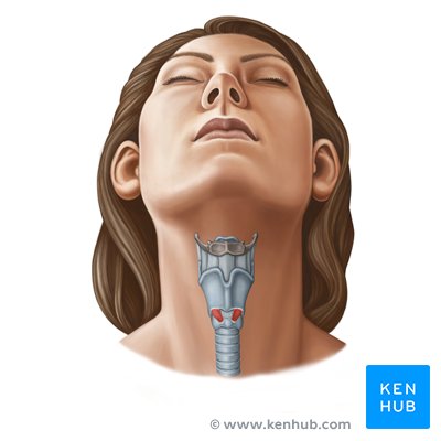
- Thyroid Gland: This butterfly-shaped endocrine gland, located anteriorly in the neck, produces hormones vital for metabolism, growth, and development. Closely associated are the parathyroid glands, which regulate calcium levels in the blood.
- Lymphatic System: The neck is rich in lymph nodes, forming extensive chains (e.g., submental, submandibular, cervical, supraclavicular). These nodes are critical components of the immune system, filtering lymph and playing a significant role in detecting and fighting infections and cancers.
Practical Tip: Given the density of critical structures, any trauma, infection, or pathological growth in the neck necessitates careful evaluation due to the potential for widespread functional impairment.
The Neck's Protective Layers: Cervical Fascias
The neck's vital contents are not just randomly arranged but are strategically organized and protected by layers of connective tissue known as cervical fascias. These fascias compartmentalize the neck, providing support, facilitating movement between structures, and importantly, limiting the spread of infection. While the reference context highlights the superficial cervical fascia, it's essential to understand the three main deep cervical fascias:
- Investing (Superficial) Fascia: This outermost layer of the deep cervical fascia completely encircles the neck, enveloping the sternocleidomastoid and trapezius muscles. It extends from the base of the skull to the clavicles and sternum.
- Pretracheal Fascia: Located anteriorly, this fascia surrounds the visceral structures of the neck, including the thyroid gland, trachea, and esophagus. It helps contain these organs and can guide the spread of infections to the mediastinum.
- Prevertebral Fascia: Posteriorly, this fascia encloses the vertebral column and the deep muscles of the neck (prevertebral and scalene muscles). It forms a protective sheath for the spinal cord and can direct infections upwards to the cranial base or downwards into the thorax.
These fascial layers are clinically significant, as they dictate the potential pathways for infections or fluid collections within the neck, guiding surgical approaches and diagnostic considerations.
In conclusion, the neck truly embodies the concept of a crossroads. Its intricate Anatomia Del Cuello reveals a remarkable density of vessels, nerves, and visceral pathways, all expertly bundled and protected within a dynamic muscular and skeletal framework. From sustaining basic life functions like breathing and swallowing to enabling complex movements and communication, the neck's role is indispensable. Its complexity makes it a fascinating area of study and a region of critical clinical importance, where understanding each component is key to appreciating the whole.