Unveiling the Neck's Intricate Anatomy: A Journey Through Musculature
The human neck, a seemingly simple structure supporting our head, is in fact a marvel of biological engineering. Far from being a mere pillar, it's a dynamic, flexible conduit packed with an astounding array of muscles, vessels, nerves, and visceral pathways, all working in concert. Understanding the
anatomia del cuello (anatomy of the neck) is crucial, not only for medical professionals but for anyone seeking to comprehend the mechanics behind head movement, speech, swallowing, and even respiration. This comprehensive exploration delves deep into the neck's complex musculature, from the superficial layers to the deep stabilizers, highlighting their functions and clinical significance.
Defining the Neck's Architecture: Boundaries and Support
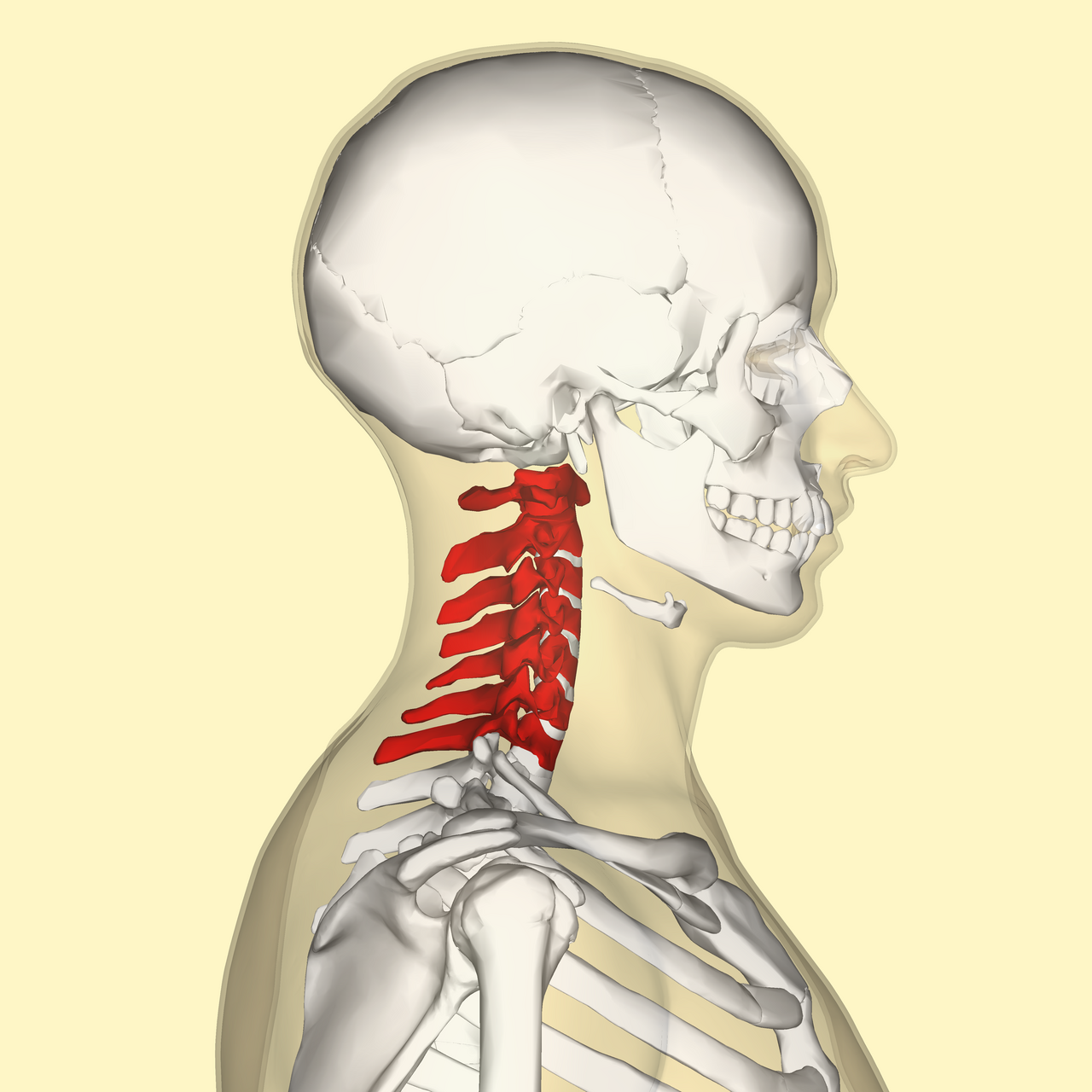
The neck serves as a vital transition zone, bridging the head with the trunk and upper limbs. Its overall shape is typically described as cylindroid, though its morphology can vary considerably based on factors like age, sex, and individual build. From an anatomical perspective, its length is determined by the superimposition of the seven cervical vertebrae (C1-C7), each with unique characteristics that allow for extensive movement. C1 (atlas) and C2 (axis) are particularly specialized, facilitating nodding and rotation. The width and thickness of the neck are influenced by the development of its substantial muscle masses and subcutaneous fatty tissue.
Its boundaries are precisely defined:
- Superior Limit: An imaginary line extending from the chin (mentum) anteriorly, along the inferior border of the mandible, past the angle of the mandible, up to the mastoid process behind the ear, and finally to the external occipital protuberance posteriorly. This line encompasses important structures like the submandibular gland, submental and submandibular lymph nodes, and parts of the facial artery and vein.
- Inferior Limit: Formed by the superior aspect of the clavicles, the superior border of the manubrium of the sternum, and a line connecting the acromial processes to the spinous process of the 7th cervical vertebra (C7).
- Posterior Limit: A convergence point involving the sternocleidomastoid, splenius capitis, and levator scapulae muscles. Notably, Erb's point, where the greater auricular nerve emerges, is a key landmark in this region.
- Anterior Limit: Defined by the midline confluence of the common fascia and the pre-laryngeal muscles, often referred to as the infrahyoid "white line."
This intricate framework provides protection and passage for critical elements, making the neck a true crossroads of the body. To learn more about these vital pathways, consider exploring our detailed article on
Neck as a Crossroads: Vessels, Nerves, & Visceral Pathways.
The Core Musculature: A Deep Dive into Neck Regions
The muscles of the neck are broadly categorized into three main regions: anterior (hyoid), lateral, and prevertebral. Each group plays a distinct, yet interconnected, role in the neck's multifaceted functions.
Suprahyoid Muscles: Orchestrators of Swallowing and Speech
Positioned superior to the hyoid bone, these four muscles form the floor of the mouth and are indispensable for vital functions like swallowing (deglutition) and speech articulation. Their coordinated action elevates the hyoid bone and, consequently, the larynx, or depresses the mandible.
- Digastric Muscle: Comprising an anterior and posterior belly, it's crucial for elevating the hyoid bone during swallowing and depressing the mandible (opening the mouth).
- Stylohyoid Muscle: Originating from the styloid process of the temporal bone, it elevates and retracts the hyoid bone, further aiding in the swallowing process.
- Mylohyoid Muscle: Forming the muscular floor of the mouth, this muscle elevates the hyoid bone and the tongue, essential for pressing food against the palate during chewing and initiating swallowing.
- Geniohyoid Muscle: Lying superior to the mylohyoid, it pulls the hyoid bone anteriorly and superiorly, further contributing to tongue movement and swallowing.
Infrahyoid Muscles: Stabilizers of the Hyoid and Larynx
Located inferior to the hyoid bone, these "strap muscles" are vital for depressing the hyoid bone and the larynx after their elevation during swallowing. They also stabilize the hyoid, providing a base for the suprahyoid muscles to act upon.
- Sternohyoid Muscle: Extends from the sternum to the hyoid, primarily responsible for depressing the hyoid bone.
- Omohyoid Muscle: Uniquely featuring two bellies (superior and inferior) connected by an intermediate tendon, it depresses and retracts the hyoid, also helping to open the cervical fascial spaces.
- Sternothyroid Muscle: Runs from the sternum to the thyroid cartilage, depressing the larynx.
- Thyrohyoid Muscle: Connects the thyroid cartilage to the hyoid bone, it can either depress the hyoid or, if the hyoid is fixed, elevate the larynx.
Lateral Neck Muscles: Dynamic Movers and Respiratory Aids, Focusing on Scalenes
The lateral region of the neck houses powerful muscles responsible for head movement and, in the case of the scalenes, accessory respiration.
- Platysma (Cutaneous Muscle of the Neck): A thin, superficial muscle that contributes to facial expressions (pulling down the corners of the mouth) and slightly depresses the mandible.
- Sternocleidomastoid (SCM) Muscle: A prominent, easily palpable muscle running from the sternum and clavicle to the mastoid process. Its unilateral contraction rotates the head to the opposite side and laterally flexes it to the same side. Bilateral contraction flexes the neck (e.g., nodding). It also plays an accessory role in respiration. The SCM forms key anatomical landmarks, dividing the neck into anterior and posterior triangles.
- Scalene Muscles (Anterior, Middle, and Posterior): These three muscles originate from the cervical vertebrae and insert onto the first and second ribs.
- Actions: Unilaterally, they contribute to lateral flexion of the neck. Bilaterally, they assist in neck flexion. Crucially, they elevate the first two ribs during forced inspiration, acting as accessory respiratory muscles.
- Clinical Significance: The space between the anterior and middle scalene muscles, known as the interscalene triangle, is a critical passage for the brachial plexus (nerves to the arm) and the subclavian artery. Compression in this region can lead to Thoracic Outlet Syndrome (TOS), causing pain, numbness, and weakness in the upper limb.
- Rectus Lateralis Capitis: Small, deep muscle for lateral flexion of the head.
Prevertebral Muscles: Deep Stabilizers of the Cervical Spine
These muscles lie directly anterior to the cervical vertebrae and are vital for stabilizing the neck and subtle head movements.
- Rectus Capitis Anterior & Longus Capitis: Primarily responsible for flexion of the head at the atlanto-occipital joint.
- Longus Colli Muscle: A long, flat muscle spanning multiple cervical and thoracic vertebrae, it contributes to flexion and subtle rotation of the cervical spine.
Beyond Muscles: The Neck's Integral Components
While our focus is on musculature, a comprehensive understanding of the
anatomia del cuello requires acknowledging other critical structures intertwined with these muscles. The neck serves as a complex conduit for the spinal cord within the cervical vertebral column. It houses major arteries (e.g., carotid arteries) and veins (e.g., jugular veins), an extensive network of lymph nodes vital for immune function, and essential visceral organs like the pharynx (involved in swallowing), larynx (voice box), trachea (windpipe), and the thyroid gland (endocrine function). Overlying and compartmentalizing these structures are the cervical fascias – superficial, pretracheal, and prevertebral – which provide support, protection, and allow for frictionless movement. Further details on these intricate structures and their functions can be found in our article:
Unveiling Neck Anatomy: Structure, Muscles, & Essential Limits.
Maintaining Neck Health: Practical Insights
Given the neck's profound complexity and its role in nearly every aspect of daily life, maintaining its health is paramount.
- Posture Awareness: Poor posture, especially "forward head posture" common with prolonged computer or phone use, can significantly strain the suprahyoid, infrahyoid, and particularly the deep cervical flexors and extensors, leading to chronic pain and dysfunction. Aim for an upright posture with the ears aligned over the shoulders.
- Ergonomics: Adjust your workstation, monitor height, and chair to support a neutral neck position.
- Regular Movement and Stretching: Gentle neck stretches and rotations can improve flexibility and blood flow to the muscles. Think about controlled movements that engage the SCM, scalenes, and deep cervical muscles.
- Strengthening Exercises: Targeted exercises, often guided by a physical therapist, can strengthen the deep neck flexors and extensors, which are crucial for stability and preventing injury.
- Listen to Your Body: Persistent neck pain, stiffness, or symptoms radiating into the arms (which could indicate nerve compression involving the brachial plexus near the scalenes) should prompt a consultation with a healthcare professional.
Understanding the complex interplay of these muscles – how the suprahyoids elevate, infrahyoids depress, the SCM rotates, and the scalenes assist in breathing – empowers us to appreciate the neck's resilience and vulnerability.
Conclusion
The neck is undeniably a masterpiece of biological design, integrating structure, function, and protection within a relatively small area. From the delicate, coordinated actions of the suprahyoid and infrahyoid muscles crucial for speech and swallowing, to the powerful rotational capabilities of the sternocleidomastoid, and the subtle yet critical respiratory assistance and nerve pathways facilitated by the scalenes, every muscle contributes to its remarkable versatility. A deeper appreciation for the
anatomia del cuello not only enriches our biological understanding but also equips us with the knowledge to better care for this vital and often overworked region of the body.